Last week, in collaboration with the SpecLab, I had the privilege of inviting MELT (Ren Loren Britton + Iz Paehr) and Shauna Janssen from Concordia University for a three-day workshop/gathering at the Kunstgewerbemuseum, with Claudia Banz as our host. The vibrant group of participants and the museum's cosy atmosphere created a splendid contrast against its spacious and brutalist architecture.
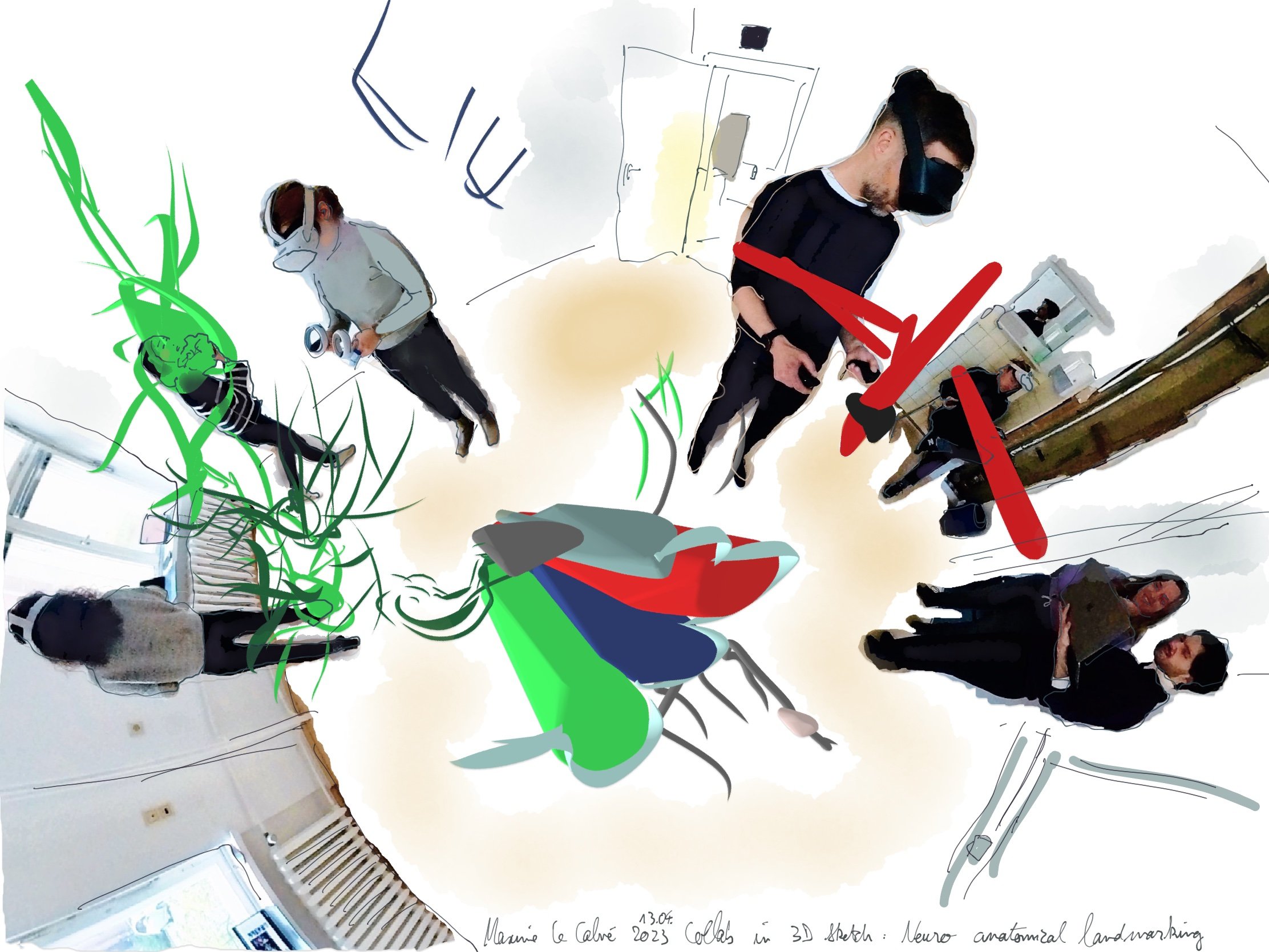
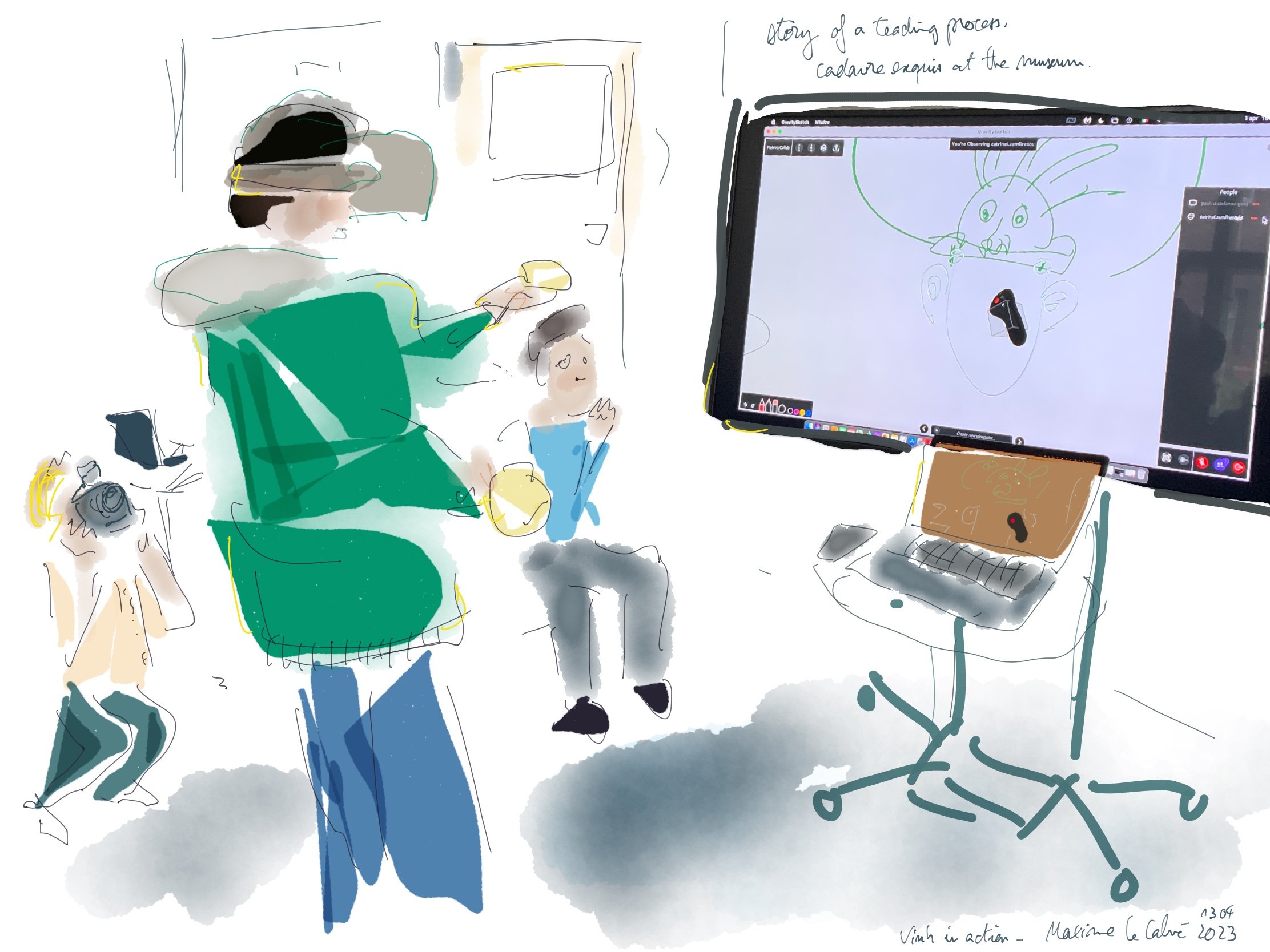
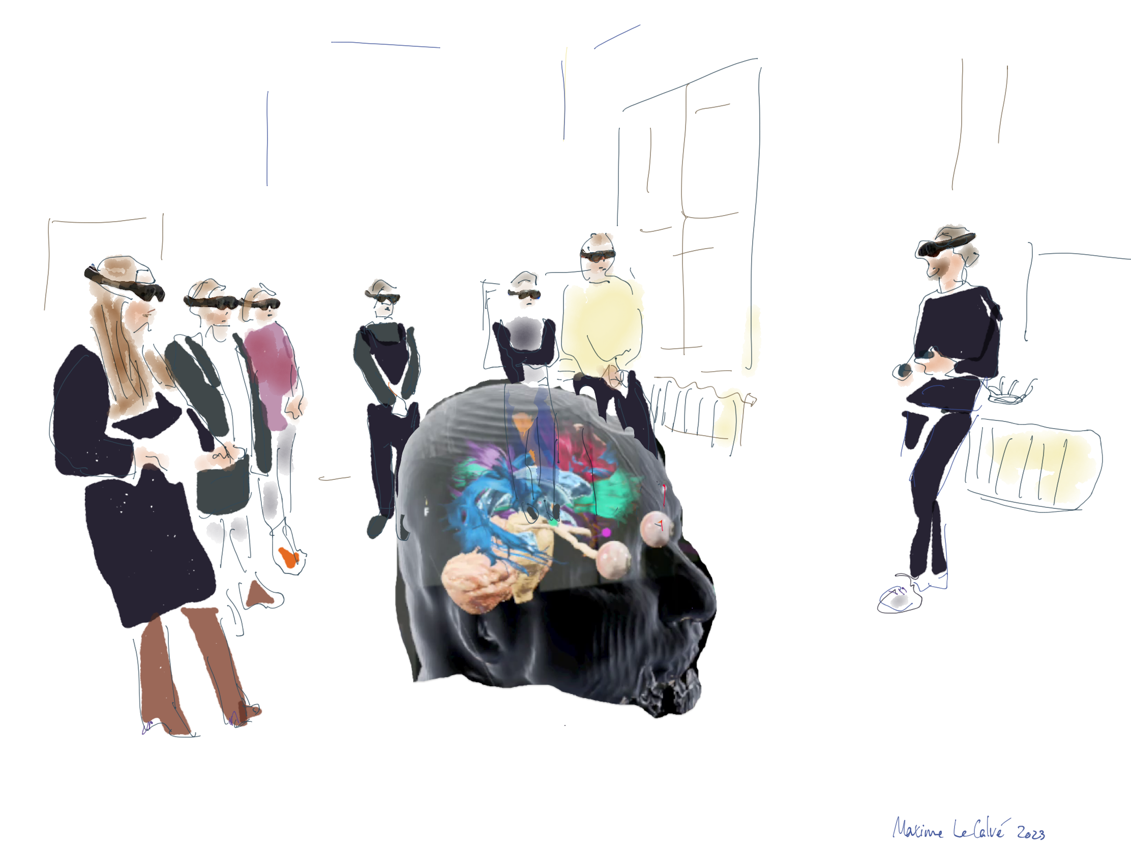
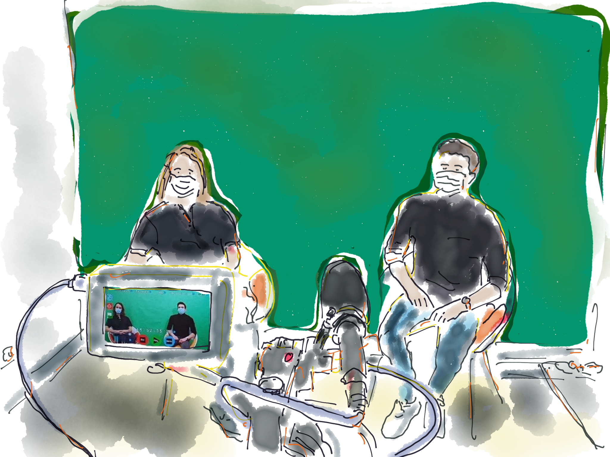
Ren and Iz led the morning sessions, while Shauna unfortunately had to remain at home. However, her insights on “Site Writing” continued to resonate with us throughout the workshop. In the afternoons, the SpecLab team presented multiple resources related to our ongoing research on brain data navigation at Charité. The focal point of this presentation was the early-stage prototype itself, which can be seen in the image above, as neuroscientist Melina Engelhardt interacts with the data.

MELT introduced us to their tools, such as the Collective Conditions, an exercise called "Warming Up to Theory," in which we danced several texts that they had selected, and the "Rituals Against Barriers."

On the first day, we used "frottage" to connect with the area around us. Inspired by Shauna's guidance, we continued with "frottage" and rubbings using technologies like Lidar and photogrammetry scanning apps. This allowed us to rub objects that are usually kept behind glass, like those in display cases. We imported these rubbings into mixed reality glasses and continued rubbing with them.
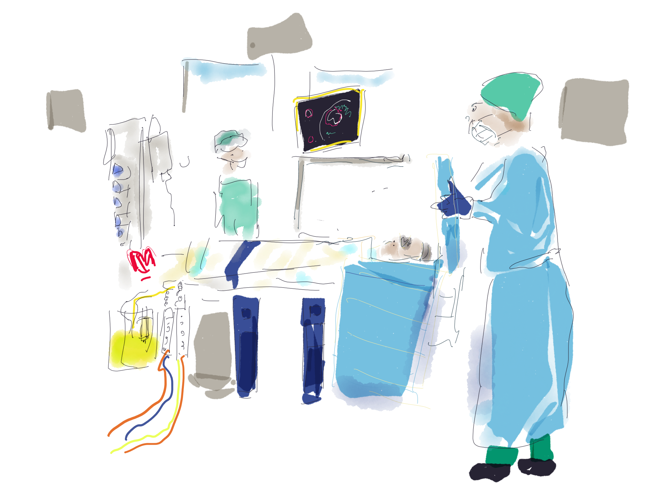
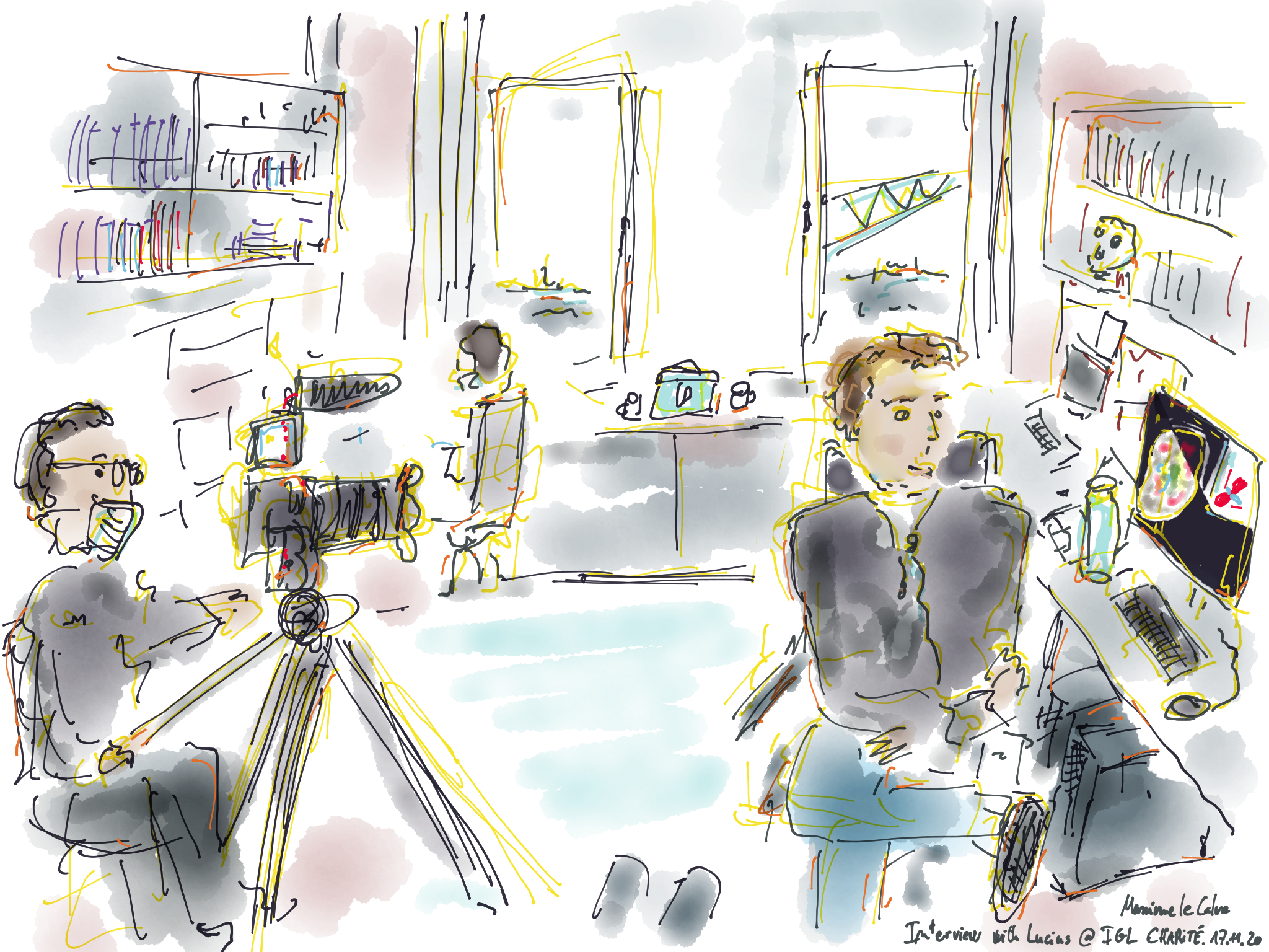
On the second day, after speaking with neurosurgeon Thomas Picht and our colleagues from the Image Guidance Lab at Charité, we started thinking about brain data and our neurosurgical case study in terms of rubbing. How can we effectively rub with the spatial information to better understand it? We had a productive and relaxing time, and we're eagerly anticipating our next workshops at the Kunstgewerbemuseum. We'll be exploring how to engage visitors with the material processes and the enjoyable work of craft people through immersive installations and score. Stay tuned for more on the "Quasi-Makers" series event at KGM.

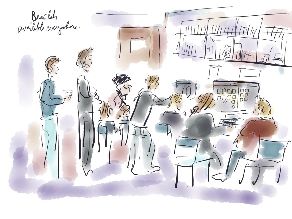
Claudia, Lin & Ren enjoying the results of the rubbing during the final public workshop session on the Friday afternoon.

Neuroscientists Melina Engelhardt and Robert Schenk grasping for brain tracts in our prototype at KGM, assisted by creative coder Warja Rybakova ©Maxime Le Calvé