The STS-ing Conference had a bold theme this year: Before Ruins. The event of the youngest professional network of Science and Technology Studies scholars in Germany was organized by the RUST Lab at the Ruhr University in Bochum.
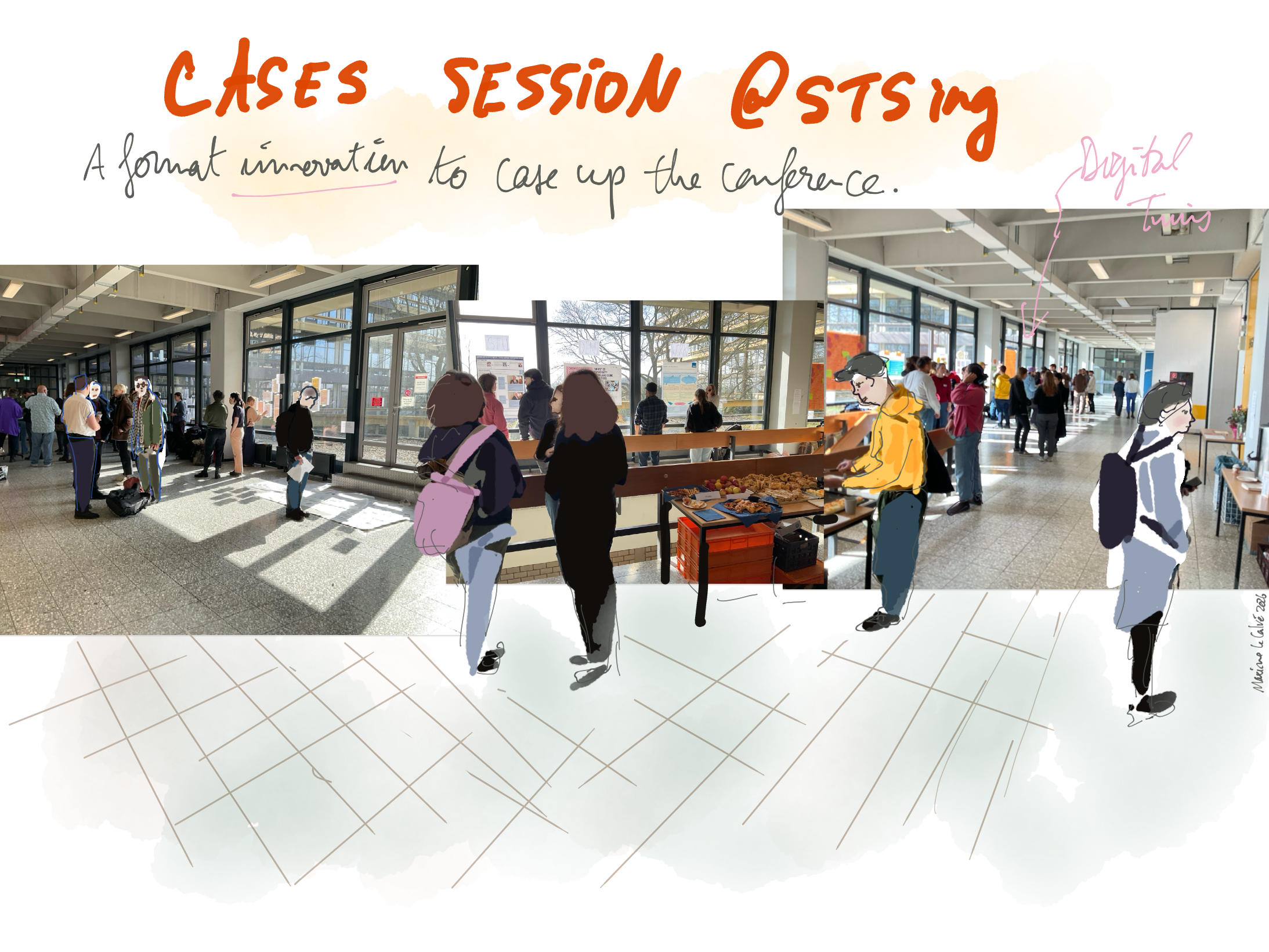
True to the highly reflective and experimental spirit of the discipline, they centered the event on a “case-study” day. Many of these groups went out for a field trip, and reported a fun and warm day despite near glacial temperatures.
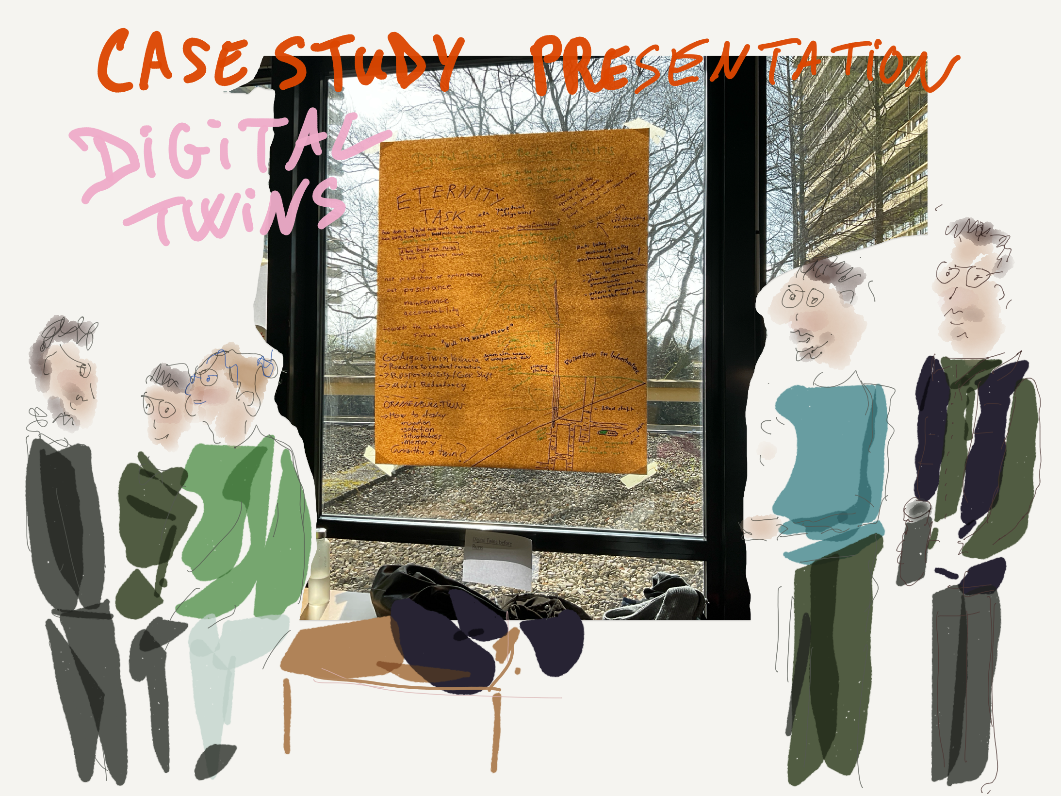
I registered with the “Digital Twin” case study with Randi Heinrich and Armin Bevergungen. While I couldn’t join the tour (we are caring for actual twins at home), they visited of the RAG Pluto mining facilities, which nowadays operates as a water-pumping institution to mitigate the legacy of a century of coke mining. “An eternity task.”
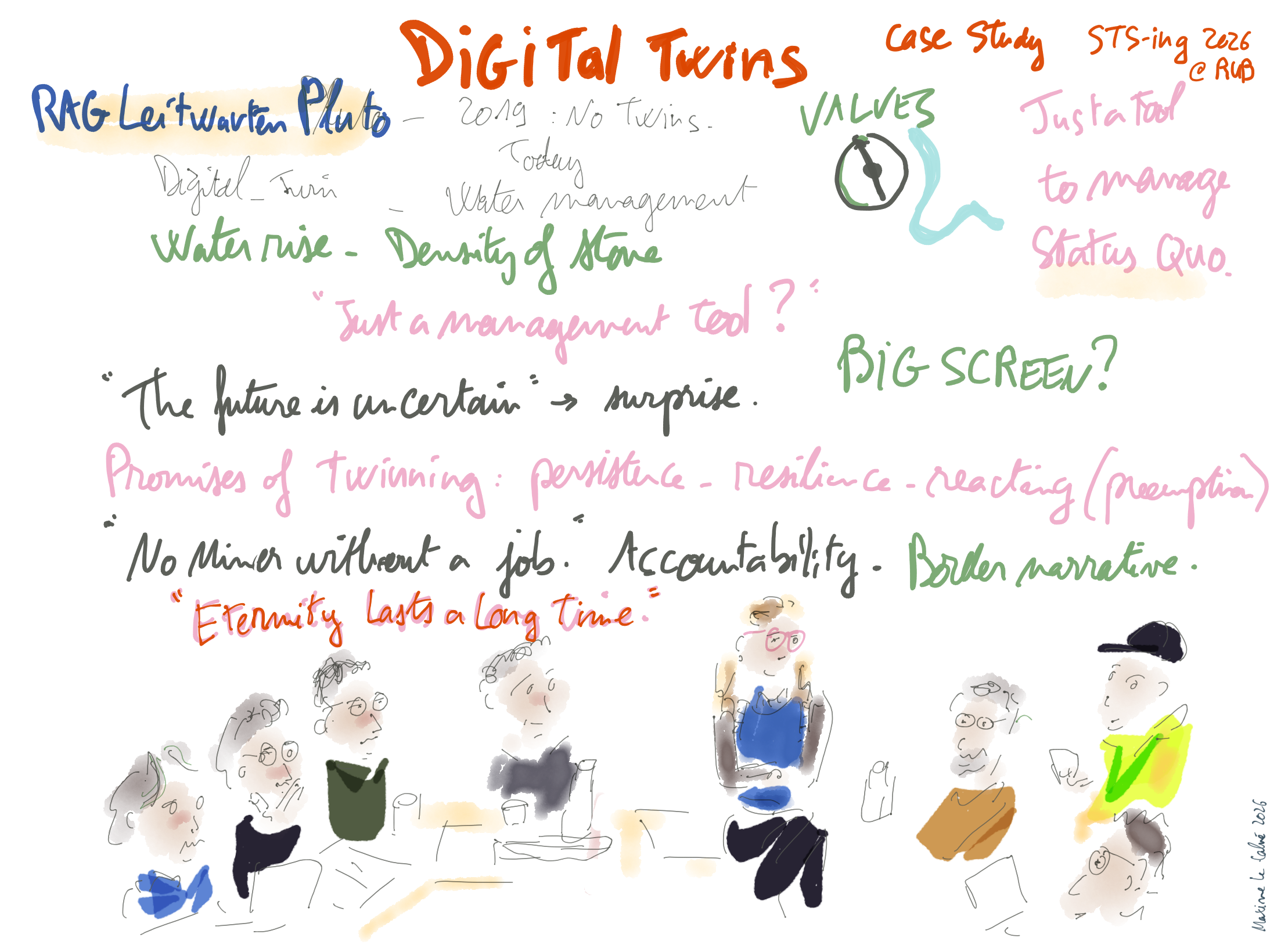
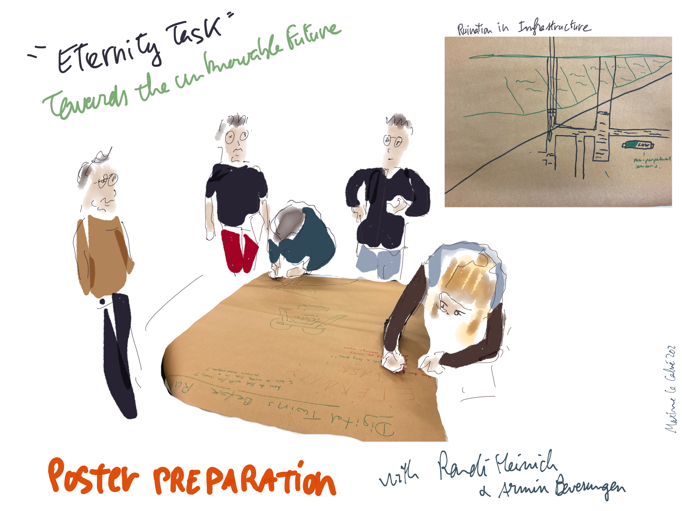
Preparing the poster together
The Bochum STS-ing team prompted us to collaborate together with strangers in a relatively informal and low pressure environment, a solid take on the challenging and competitive atmosphere of most of these events.
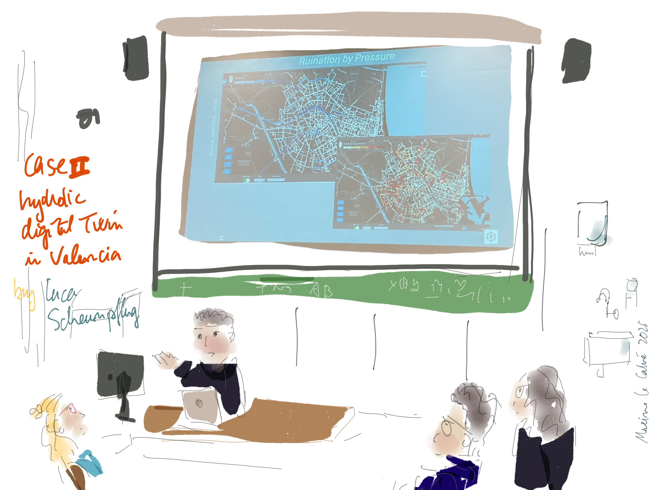
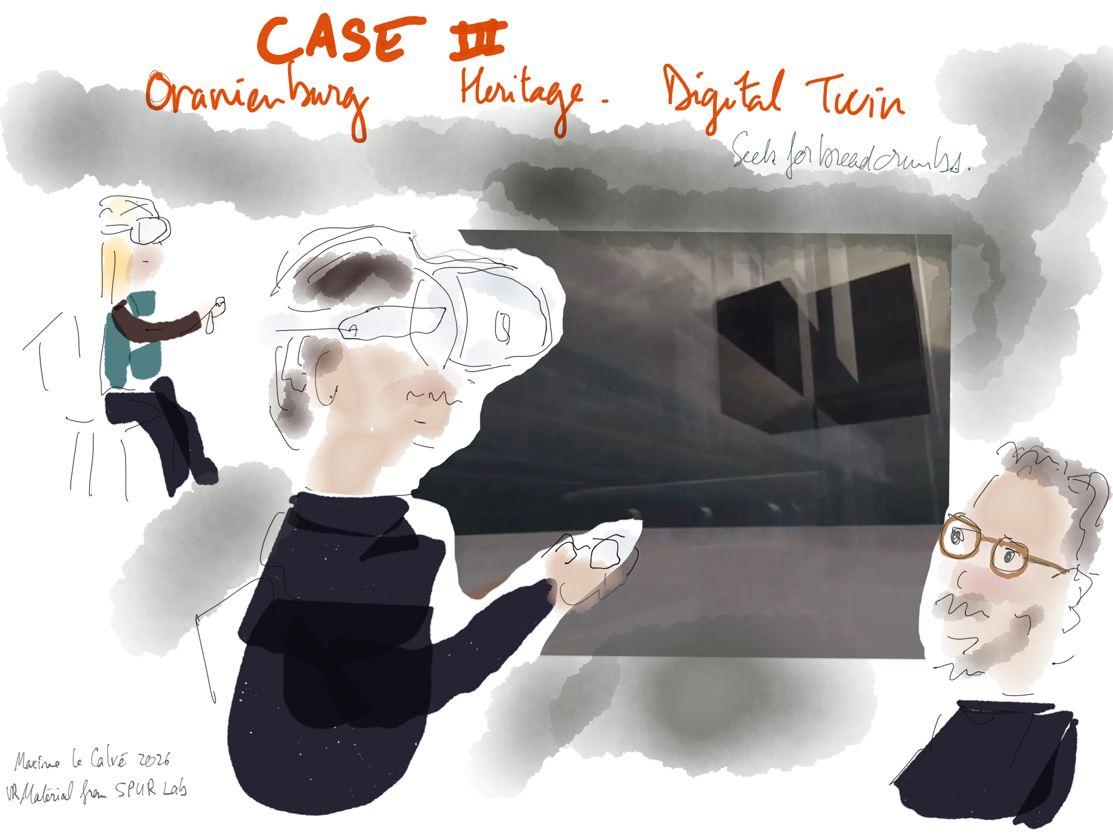
Additionally to the mining legacy case, two other participants presented their work on diverse digital twinning: Luca Scheunpflug on his coming fieldwork in Valencia’s hydraulic systems. Then Jens Fehrenbacher on digital twins of heritage places in virtual reality, with the SPUR-Lab reconstruction of Oranienburg concentration camp.
The next morning, in a big space flooded with much welcome sun, the case study presentation was a highlight. A very relaxed moment that served as the perfect warm up for the paper sessions.
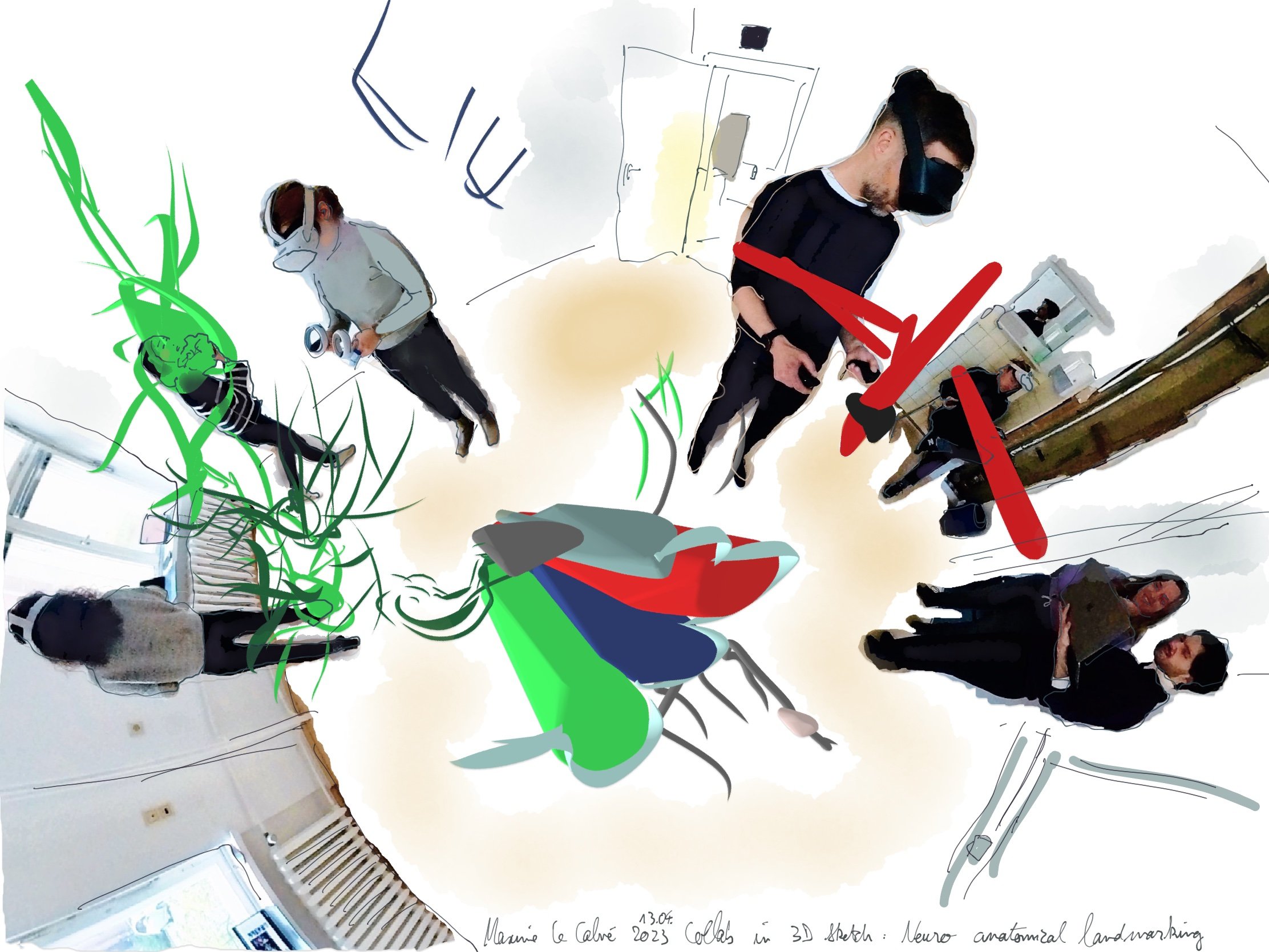
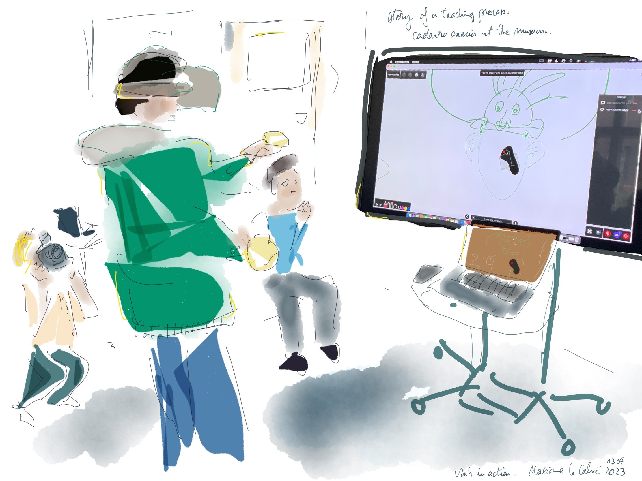
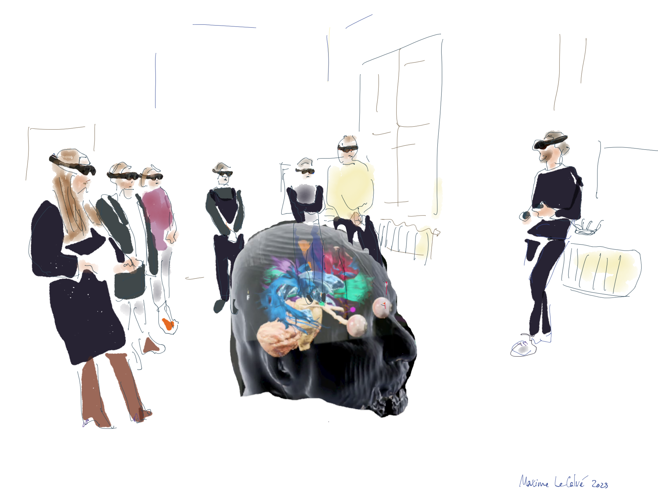
Speaking of digital twins, I was delighted to present new images from our prototype developed at the Speculative Realities Lab at the Charité Berlin. My paper “Braining the Critical Zone: Neurosurgery and the Logic of Digital Twins” discussed the totalizing endeavour of tractographic brain modelling and pitted it against our own experimental attempts to make diffusion data experience-able, refraining from “feigning certainty” (Amoore 2025).
Before the ruins, we need to develop new ways of dwelling in the critical zone.
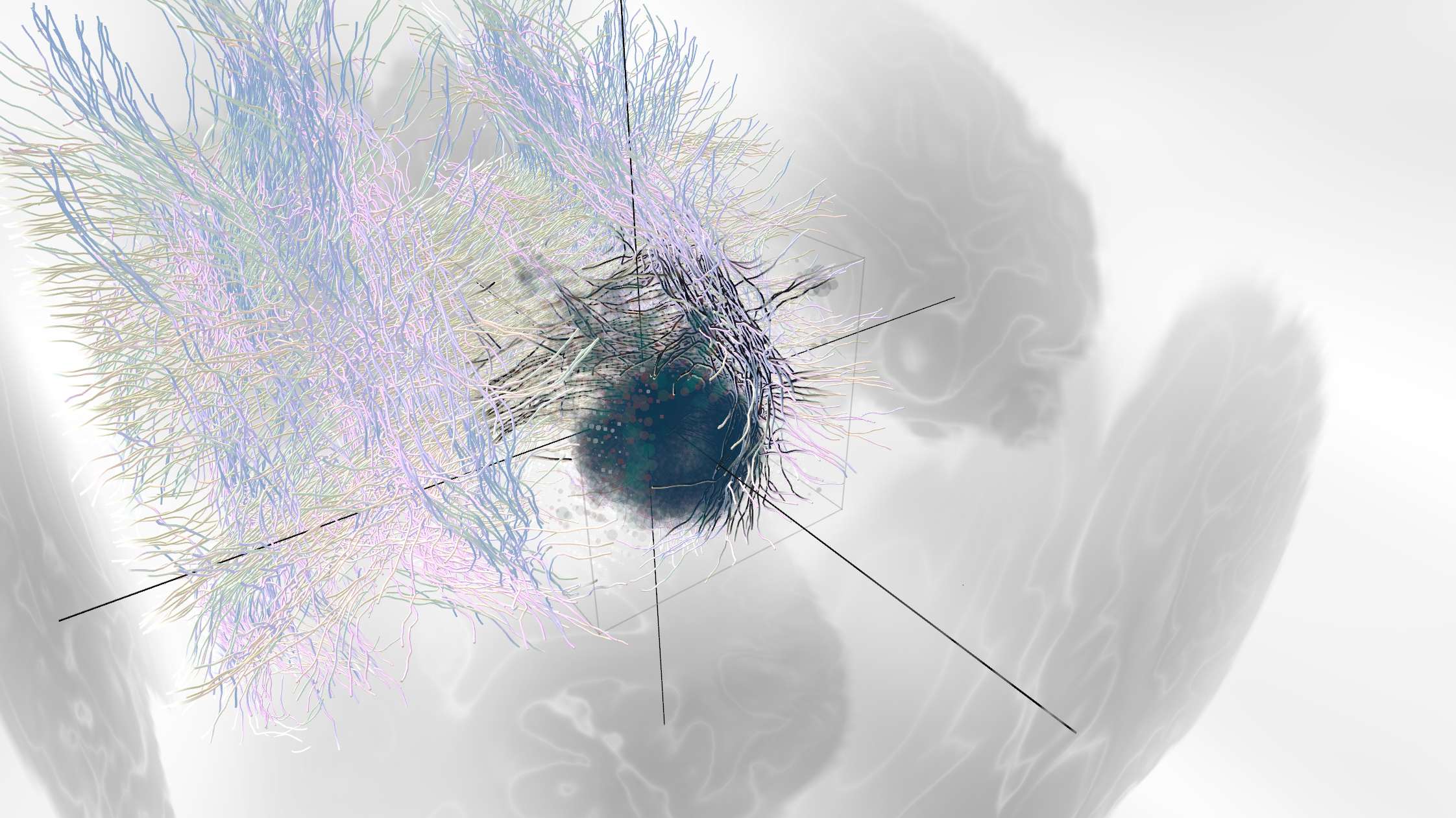
Topovox protoype, developed by the Speculative Realities Lab
Many thanks to the RUST Lab team for this amazing event, and to Randi and Armin for the Digital Twin case.